

The PAMCET Learning Team is the institutional curriculum and content group behind course publishing on this platform. PAMCET (Professional and Academic Management Centre for Education and Training) is operated by Digitpen Hub Ltd, a company registered in Lagos, Nigeria. Courses credited to the PAMCET Learning Team are structured, organized, and published centrally by the platform rather than authored by a single named individual instructor — each course's syllabus, learning outcomes, and content are reviewed before publication under this institutional account. For questions about a specific course or to report a content issue, please use the platform's Help & Support or Contact Us pages.
B.Sc. DegreeFaculty of Faculty of Medical, Health & Clinical SciencesRadiography
Introduction to Radiography
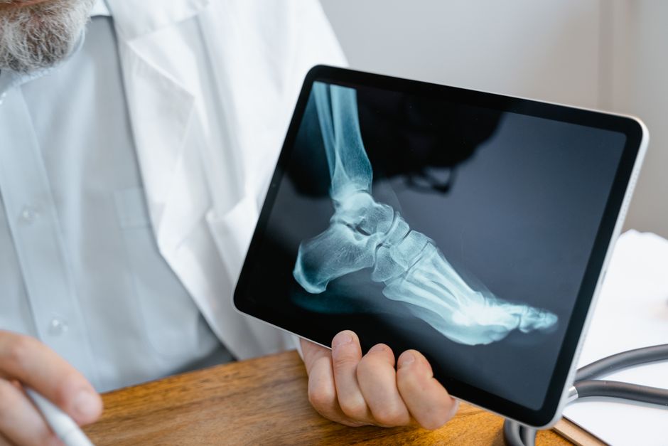
An introductory course covering fundamental principles of Radiography.

Course Overview
An introductory course covering fundamental principles of Radiography. Curriculum Structure & Core Modules: • 1. Introduction to Introduction to Radiography & Core Frameworks • 2. Theoretical Foundations & Foundational Models of Introduction to Radiography • 3. Advanced...
Degree Programme Curriculum Context
This course is a curriculum unit of the B.Sc. Radiography degree framework in Nigeria.
Earn Your Certificate
Showcase your skills with a certificate! Complete the course, submit your project, and earn your certificate. Here's a sample of what you'll receive to show off your accomplishments.

Lecture Board & Syllabus
Course Curriculum
Browse complete theoretical foundations, worked calculations, laboratory protocols, and board assessment questions under each topic.
8 Chapters
•24 Lecture
•0s
Subtopic 1Document Lecture
Introduction to Introduction to Radiography & Core Frameworks - Concept Explanation & Foundations
Section 1: Detailed Theoretical Foundations & Conceptual Framework The discipline of diagnostic radiography and medical imaging originated on November 8, 1895, when German physicist Wilhelm Conrad Röntgen discovered an unknown form of penetrating electromagnetic radiation while experimenting with Crookes and Hittorf gas-discharge cathode ray tubes at the University of Würzburg. Observing the green...
Module 1 • Subtopic 1
Subtopic 2Document Lecture
Introduction to Introduction to Radiography & Core Frameworks - Worked Examples & Case Analysis
Section 1: Detailed Theoretical Foundations & Conceptual Framework The formation of a diagnostic medical image depends entirely upon the differential attenuation of X-ray photons as they traverse biological tissues of varying atomic compositions, physical densities, and thicknesses. Attenuation is defined as the total reduction in the intensity of an X-ray beam as it traverses matter, resulting fr...
Module 1 • Subtopic 2
Subtopic 3Document Lecture
Introduction to Introduction to Radiography & Core Frameworks - Practice Questions & Exercises
Section 1: Detailed Theoretical Foundations & Conceptual Framework Modern diagnostic radiographic equipment comprises an integrated electromechanical system engineered to generate, modulate, restrict, and capture X-ray photon beams with high spatial precision. The X-ray tube housing incorporates lead lining to attenuate isotropic leakage radiation to regulatory limits (< 1.0 mGy/hour at 1.0 met...
Module 1 • Subtopic 3
Subtopic 1Document Lecture
Theoretical Foundations & Foundational Models of Introduction to Radiography - Concept Explanation & Foundations
Section 1: Detailed Theoretical Foundations & Conceptual Framework The technological evolution of medical image receptors over the past century represents a progressive transition from chemical photochemical emulsion film systems to digital solid-state matrix detectors. The foundational architecture of classical screen-film radiography relied upon Silver Halide Emulsions (silver bromide AgBr and s...
Module 2 • Subtopic 1
Subtopic 2Document Lecture
Theoretical Foundations & Foundational Models of Introduction to Radiography - Worked Examples & Case Analysis
Section 1: Detailed Theoretical Foundations & Conceptual Framework The creation of a diagnostically efficacious medical radiograph demands meticulous manipulation of physical exposure parameters and geometric factors to optimize image quality. In classical and digital radiography, image quality is governed by four cardinal photographic and geometric properties: Receptor Exposure (Density): The tot...
Module 2 • Subtopic 2
Subtopic 3Document Lecture
Theoretical Foundations & Foundational Models of Introduction to Radiography - Practice Questions & Exercises
Section 1: Detailed Theoretical Foundations & Conceptual Framework Contrast media represent specialized diagnostic pharmacological agents administered to temporarily alter the radiodensity of internal biological cavities, vascular lumens, and gastrointestinal tracts, thereby accentuating subject contrast between structures with near-identical native linear attenuation coefficients. Radiographic co...
Module 2 • Subtopic 3
Subtopic 1Document Lecture
Advanced Concepts, Variables & Mechanics - Concept Explanation & Foundations
Section 1: Detailed Theoretical Foundations & Conceptual Framework Radiation protection and radiobiology form the ethical and scientific foundation of professional radiographic practice. Ionizing radiation possesses sufficient quantum energy to eject orbital electrons from biological atoms, initiating a cascade of chemical bond ruptures, molecular lesions, and cellular damage. The radiobiological...
Module 3 • Subtopic 1
Subtopic 2Document Lecture
Advanced Concepts, Variables & Mechanics - Worked Examples & Case Analysis
Section 1: Detailed Theoretical Foundations & Conceptual Framework Radiographic positioning represents the meticulous clinical art and science of spatially orienting the human body and directing the central X-ray beam to demonstrate specific anatomical structures with diagnostic fidelity. Human anatomy is three-dimensional; however, conventional projection radiography compresses this volume into a...
Module 3 • Subtopic 2
Subtopic 3Document Lecture
Advanced Concepts, Variables & Mechanics - Practice Questions & Exercises
Section 1: Detailed Theoretical Foundations & Conceptual Framework Fluoroscopy represents dynamic, real-time functional radiographic imaging utilized to visualize moving physiological processes (cardiac wall motion, vascular hemodynamics, peristalsis, and deglutition) and guide percutaneous interventional procedures. Invented by Thomas Edison in 1896 utilizing calcium tungstate phosphor screens, e...
Module 3 • Subtopic 3
Subtopic 1Document Lecture
Methodological Approaches & System Analysis - Concept Explanation & Foundations
Section 1: Detailed Theoretical Foundations & Conceptual Framework Computed Tomography (CT), developed independently by British electrical engineer Sir Godfrey Hounsfield and South African-American physicist Allan Cormack in the early 1970s (earning them the 1979 Nobel Prize in Physiology or Medicine), revolutionized medical diagnostics by generating cross-sectional tomographic slices of human ana...
Module 4 • Subtopic 1
Subtopic 2Document Lecture
Methodological Approaches & System Analysis - Worked Examples & Case Analysis
Section 1: Detailed Theoretical Foundations & Conceptual Framework Magnetic Resonance Imaging (MRI) represents a non-ionizing, multi-planar diagnostic imaging modality utilizing nuclear magnetic resonance (NMR) phenomena to produce soft-tissue anatomical detail. Discovered independently by Felix Bloch and Edward Purcell in 1946 (earning them the 1952 Nobel Prize in Physics), NMR was transformed in...
Module 4 • Subtopic 2
Subtopic 3Document Lecture
Methodological Approaches & System Analysis - Practice Questions & Exercises
Section 1: Detailed Theoretical Foundations & Conceptual Framework Diagnostic medical ultrasound (sonography) represents an interactive, non-ionizing, non-invasive imaging modality utilizing high-frequency acoustic pressure waves to generate real-time anatomical images and hemodynamic blood flow data. Unlike electromagnetic modalities (X-rays, CT, MRI), ultrasound waves are mechanical longitudinal...
Module 4 • Subtopic 3
Subtopic 1Document Lecture
Analytical Case Studies & Research Applications - Concept Explanation & Foundations
Section 1: Detailed Theoretical Foundations & Conceptual Framework Systematic radiographic image interpretation and pathology recognition represent vital clinical competencies for diagnostic radiographers and medical imaging practitioners. In emergency and critical care environments, the ability to rapidly identify acute, life-threatening radiological abnormalities ensures immediate interventional...
Module 5 • Subtopic 1
Subtopic 2Document Lecture
Analytical Case Studies & Research Applications - Worked Examples & Case Analysis
Section 1: Detailed Theoretical Foundations & Conceptual Framework Musculoskeletal radiography represents the highest-volume clinical domain in diagnostic imaging, providing essential objective data for orthopedic trauma management, fracture reduction, and deformity tracking. A fracture is defined biomechanically as a structural disruption in the continuity of cortical or trabecular bone resulting...
Module 5 • Subtopic 2
Subtopic 3Document Lecture
Analytical Case Studies & Research Applications - Practice Questions & Exercises
Section 1: Detailed Theoretical Foundations & Conceptual Framework Interventional Radiography (IR) represents a dynamic, minimally invasive subspecialty of diagnostic and therapeutic medicine utilizing real-time fluoroscopy, computed tomography, and ultrasound to perform targeted percutaneous diagnostic and therapeutic interventions. Pioneered in 1953 by Swedish radiologist Sven-Ivar Seldinger and...
Module 5 • Subtopic 3
Subtopic 4Open Access~3 min read
Mid-Semester Case Study & Research Paper
Sign in or enroll to access the full lecture notes and interactive reader.
Module 5 • Subtopic 4•115 characters of authentic academic content
Subtopic 1Document Lecture
Strategic Implementation & Policy Implications - Concept Explanation & Foundations
Section 1: Detailed Theoretical Foundations & Conceptual Framework Quality Assurance (QA) and Quality Control (QC) in medical radiography constitute the comprehensive administrative, operational, and physical testing frameworks designed to guarantee that diagnostic imaging services consistently produce images of optimal diagnostic quality while minimizing patient radiation dose and operating costs...
Module 6 • Subtopic 1
Subtopic 2Document Lecture
Strategic Implementation & Policy Implications - Worked Examples & Case Analysis
Section 1: Detailed Theoretical Foundations & Conceptual Framework Radiation protection and radiographic technique adaptation must be dynamically tailored to the extremes of human life: pediatric and geriatric patients. Pediatric patients are uniquely vulnerable to the biological effects of ionizing radiation, while geriatric patients present complex challenges related to osteoporotic skeletal dem...
Module 6 • Subtopic 2
Subtopic 3Document Lecture
Strategic Implementation & Policy Implications - Practice Questions & Exercises
Section 1: Detailed Theoretical Foundations & Conceptual Framework The operational infrastructure of modern medical imaging is entirely digital, governed by a complex, interconnected ecosystem of healthcare information systems, image archival networks, and international telecommunication communication standards. The contemporary digital imaging enterprise is structured upon four interdependent fou...
Module 6 • Subtopic 3
Subtopic 1Document Lecture
Comparative Perspectives & Modern Theories of Introduction to Radiography - Concept Explanation & Foundations
Section 1: Detailed Theoretical Foundations & Conceptual Framework Nuclear medicine and molecular imaging represent a paradigm shift in medical diagnosis: whereas conventional radiography, CT, and MRI image static structural and anatomical density distributions, nuclear medicine evaluates in vivo organ physiology, cellular biochemistry, receptor expression, and metabolic kinetics. This is accompli...
Module 7 • Subtopic 1
Subtopic 2Document Lecture
Comparative Perspectives & Modern Theories of Introduction to Radiography - Worked Examples & Case Analysis
Section 1: Detailed Theoretical Foundations & Conceptual Framework The integration of Artificial Intelligence (AI), Machine Learning (ML), and Deep Learning (DL) into medical imaging represents one of the most profound technological evolutions in radiological history. Radiography is inherently digital, quantitative, and data-dense, making it exceptionally fertile ground for automated pattern recog...
Module 7 • Subtopic 2
Subtopic 3Document Lecture
Comparative Perspectives & Modern Theories of Introduction to Radiography - Practice Questions & Exercises
Section 1: Detailed Theoretical Foundations & Conceptual Framework Evidence-Based Practice (EBP) and clinical governance in diagnostic radiography and medical imaging represent the systematic, objective framework used to ensure that imaging utilization is clinically justified, diagnostically efficacious, cost-effective, and safe. With healthcare expenditures expanding globally and medical radiatio...
Module 7 • Subtopic 3
Subtopic 1Document Lecture
Synthesis, Future Trends & Final Review - Concept Explanation & Foundations
Section 1: Detailed Theoretical Foundations & Conceptual Framework Radiographer jurisprudence, medical ethics, and patient advocacy define the legal duties, regulatory boundaries, and moral obligations governing professional radiological technologists. Diagnostic radiographers operate in high-risk clinical environments involving ionizing radiation, powerful static magnetic fields, rapid intravenou...
Module 8 • Subtopic 1
Subtopic 2Document Lecture
Synthesis, Future Trends & Final Review - Worked Examples & Case Analysis
Section 1: Detailed Theoretical Foundations & Conceptual Framework Specialized clinical radiography extends beyond standard outpatient and emergency department suites into high-stakes, unconventional operating environments: forensic medicolegal investigations, mobile bedside imaging in intensive care units, intraoperative surgical guidance, and Mass-Casualty Incident (MCI) disaster response. Radio...
Module 8 • Subtopic 2
Subtopic 3Document Lecture
Synthesis, Future Trends & Final Review - Practice Questions & Exercises
Section 1: Detailed Theoretical Foundations & Conceptual Framework The contemporary practice of medical imaging and radiography culminates in Multimodal Imaging Integration: the sophisticated, synergistic synthesis of projection radiography, computed tomography, magnetic resonance imaging, ultrasound, and nuclear medicine to solve complex, multi-system clinical dilemmas. In modern medicine, no sin...
Module 8 • Subtopic 3
Academic Ecosystem & Connected Pathways
Deepen your studies across connected learning pathways in the PAMCET academic network:
Digital Research Library16,000+ open-access monographs & textbooks
Structured Learning PathsCurated multi-course career specializations
WAEC / SSCE Examination HubComplete secondary school syllabus & mock tests
JAMB / UTME PreparationComputer-based tests & scoring analytics
Tertiary & University Hub100L - 400L degree benchmark modules
Credential Verification PortalInstant cryptographic certificate authentication
Instructor
PAMCET Learning Team
Institutional Course Curation · PAMCET
Qualifications:
PAMCET Learning Team — Institutional Course Curation, Digitpen Hub LtdMy Skills:
- Curriculum Curation
- Course Content Review
- Learning Outcome Design
- Course Publishing & Quality Assurance
- Platform Content Operations
Reviews
No reviews available
Free100% Off
Certificate available separately for $7 after completionCourse Includes
8 Chapters
24 Lectures
1 week
Skill Level beginner
Taught in English
Lifetime Access
Certification of Completion
Category: University - Radiography
Share On
Download Syllabus & Study Guide
Receive the full lecture syllabus, reading list, and milestone schedule for Introduction to Radiography.
By submitting, you agree to receive educational updates. You can unsubscribe anytime. Read our Privacy Policy.